support@rsdiagnosticcentre.in
Phone : +918147282686
EMERGENCY XRAY SERVICES IN YELAHANKA
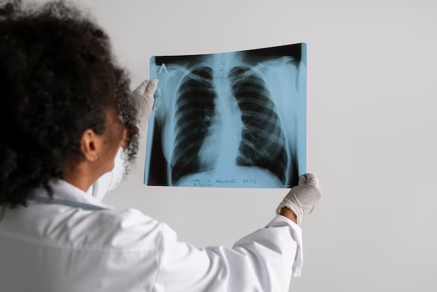
We at R S Diagnostics analyze the Lung, Heart, Blood vessels, Bones, Diaphragm and soft tissue structures to help diagnose a wide range of conditions affecting the chest and respiratory system.
1. Lungs:
The main focus of a chest X-ray is to evaluate the condition of the lungs. Radiologists look for signs of infections, such as pneumonia or tuberculosis, as well as lung diseases like emphysema or pulmonary fibrosis.
2. Heart:
The size and shape of the heart are assessed to detect any abnormalities such as enlargement or fluid around the heart. This can indicate conditions like congestive heart failure or pericarditis.
3. Blood vessels:
The X-ray can show the blood vessels in the chest, including the aorta and pulmonary arteries. Abnormalities in these vessels, such as aneurysms or pulmonary embolisms, may be visible on the X-ray.
4. Bones:
The ribs and spine are also visible on a chest X-ray. Fractures, bone tumors, and degenerative changes can be identified.
5. Diaphragm:
The diaphragm, the muscle that separates the chest cavity from the abdominal cavity, is examined for abnormalities such as paralysis or hernias.
6. Soft tissues: Other soft tissues in the chest, such as the mediastinum (the area between the lungs), are evaluated for masses or abnormal fluid collections.
Tag
XRAY SERVICES IN YELAHANKAEMERGENCY XRAY SERVICES IN YELAHANKAACCURATE XRAY SERVICES IN YELAHANKAR S DIAGNOSTIC CENTRE NEAR ANANTHPURA GATEEnquiry
Get in touch


R S DIAGNOSTIC CENTRE.All Rights Reserved © 2025